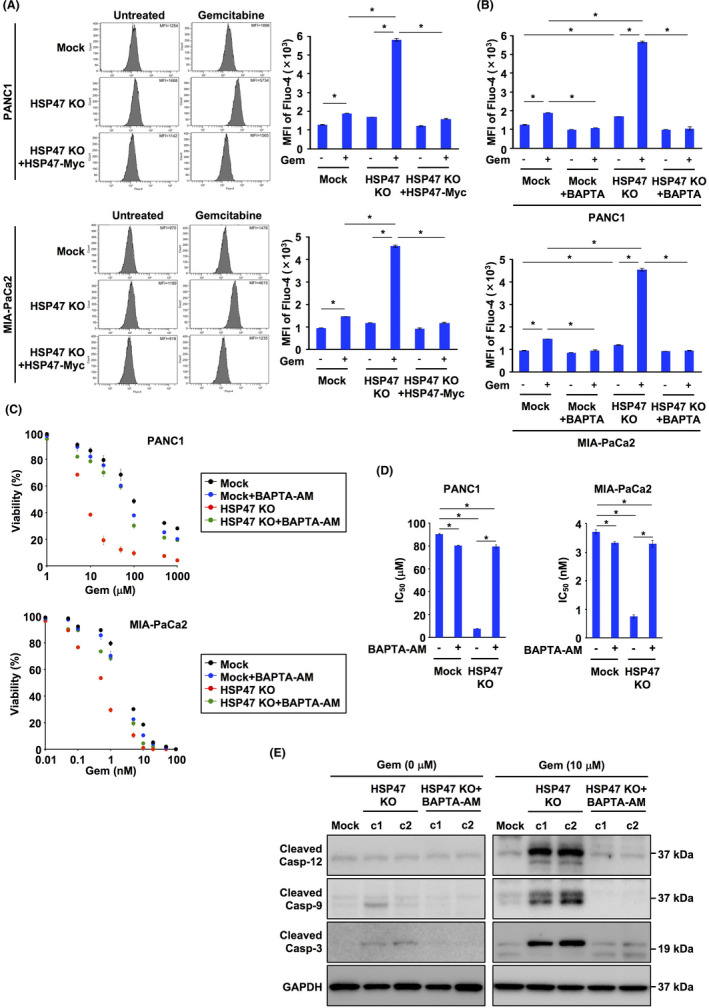
FIGURE 4.

Disruption of HSP47 induces an increase in intracellular Ca2+ levels in pancreatic cancer cells after treatment with gemcitabine. A, Levels of intracellular Ca2+ in PDAC cells (PANC1 cells and MIA‐PaCa2 cells), HSP47 KO PDAC cells (HSP47 KO PANC1 cells and HSP47 KO MIA‐PaCa2 cells) and HSP47 KO PDAC cells expressing HSP47‐Myc after treatment with gemcitabine (10 μmol/L for PANC1 cells and 1 nmol/L for MIA‐PaCa2 cells). Levels of intracellular Ca2+ were determined using flow cytometry (left panel) and calculating the intensity of Fluo‐4 (right panel). B, Levels of intracellular Ca2+ in PDAC cells and HSP47 KO PDAC cells after treatment with BAPTA‐AM (5 μmol/L) and gemcitabine. C, Viability of PDAC cells and HSP47 KO PDAC cells after treatment with BAPTA‐AM and gemcitabine. D, IC50 values of HSP47 KO PDAC cells after treatment with BAPTA‐AM and gemcitabine. E, Activation of the calcium/caspase‐12/caspase‐9/caspase‐3 axis in HSP47 KO PANC1 cells (c1 and c2) after treatment with or without BAPTA‐AM and gemcitabine. *, P < .05
