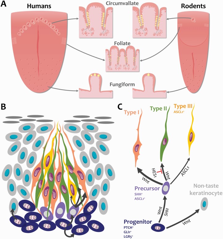
Figure 1.
Anatomy of the tongue and taste cell renewal. (A) Taste buds (yellow) are embedded in taste papillae in the tongue epithelium. The number of taste buds in fungiform papillae, which are located in the anterior two-thirds of the tongue, is species-dependent; rodent fungiform papillae each contain a single taste bud while multiple taste buds can be found in a human fungiform papilla. Hundreds of taste buds line the trenches (epithelial invaginations) of foliate and circumvallate papillae in the posterior tongue. In human circumvallate papillae, taste buds are mostly found in the inner wall of the trench. Foliate papillae lie laterally while circumvallate papillae are organized in a central V-shaped formation. Rodents possess only a single circumvallate papilla. (B) Taste buds are made of 50–100 cells that are continually replaced throughout life. Progenitor cells (dark blue) reside along the basement membrane outside taste buds and actively divide to self-renew and produce taste cells and non-taste keratinocytes (light gray) that surround taste buds. Following mitosis, taste-fated lingual progenitors enter taste buds and specify into post-mitotic SHH+ taste precursor cells (magenta). Precursor cells then differentiate into most prevalent Type I glial-like cells (tangerine), Type II sweet/bitter/umami receptor cells (green), and least common Type III sour receptor cells (yellow). (C) Fate decision is regulated by the Wnt pathway, Hedgehog, and Notch signaling. The Wnt/β-catenin pathway controls all steps of taste and non-taste cell renewal, while SHH instructs progenitors to differentiate into taste cells. Notch signaling represses Type II cell fate via HES1 and transcriptionally represses ASCL1 to control Type III taste cell differentiation. Illustrations are modified from Servier Medical Art licensed under a Creative Commons Attribution 3.0 Unported License (https://smart.servier.com/).

