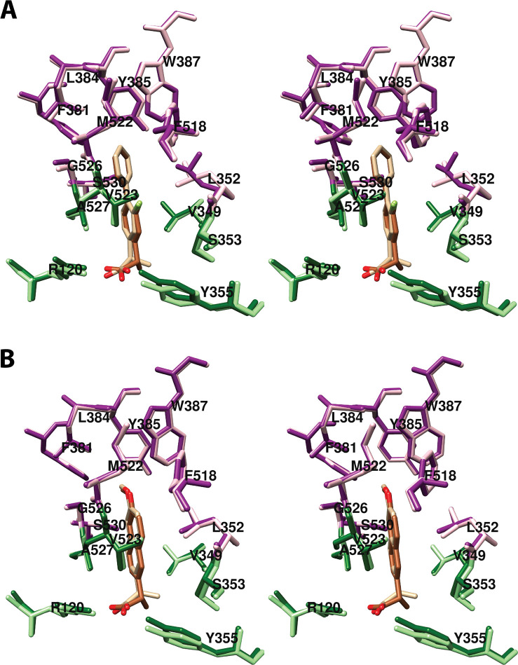
Figure 14.
Wall-eyed stereo views of overlays of the structures of (R)- and (S)-flurbiprofen (A) and (R)- and (S)-naproxen (B), bound in the cyclooxygenase active site of COX-2 and the side chains that make up the proximal binding pocket (light/dark green) and the central binding pocket (pink/magenta). In each case, structures related to the (R)-enantiomer (tan) are shown in the lighter color and those related to the (S)-enantiomer (sienna) are shown in the darker color. From PDB 3PGH and 3RR3 (A) and PDB 3NT1 and 3Q7D (B).

