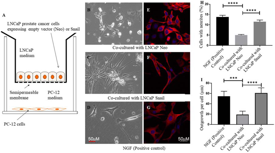
FIGURE 4. Stable overexpression of Snail in LNCaP cells promotes neuronal outgrowth in PC-12 nerve cells.

(A) LNCaP prostate cancer cells stably expressing empty vector (Neo) or Snail cDNA (LNCaP Snail) were cultured on 12-well cell culture inserts with semipermeable support membranes pre-coated with collagen type I. PC-12 cells were cultured on the bottom of these wells. Co-cultured PC-12 cells with neurite outgrowth, visualized by light microscopy (Panels B-D) were quantified based on the percentage of neuronal cells which showed a phenotype of axonal extensions. In another set of experiments, co-cultured PC-12 cells were stained with a MaP2 antibody, a dendritic marker followed by a confocal fluorescence microscopy (Panels E-G), and measurement of neurite length per cell (Panel I). Quantitated data in panels H and I are representative of 6 and 5 microscopic fields per experiment, respectively (***p< 0.001, ****p< 0.0001). Statistical analysis was done using GraphPad Prism. Nerve growth factor (NGF, 100 ng/mL) was utilized as a positive control. Scale bars are 50 μm.
