Figure 4.

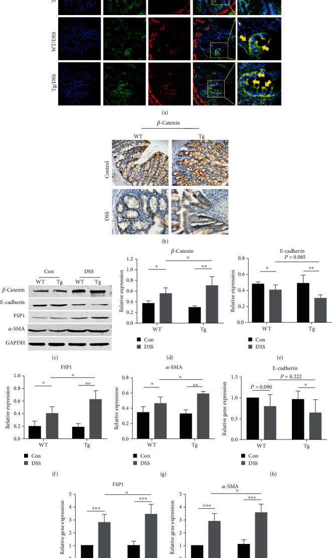
TL1A promotes EMT in the DSS-induced intestinal fibrosis model. (a) Immunofluorescence staining of colon tissue sections from wild type (WT) and transgenic (Tg) mice probed with antibodies against E-cadherin (green) and FSP1 (red) (400x). E-cadherin+FSP1+ cells (yellow; reflects EMT). Nuclei are stained with DAPI (blue). (b) Immunohistochemical staining of β-catenin in colon tissues of mice groups (400x). (c–g) Western blot analysis of total protein of β-catenin, E-cadherin, α-SMA, and FSP1 in intestinal tissue, normalized with GAPDH. (h–j) RT-PCR analysis of E-cadherin, α-SMA, and FSP1 mRNA levels in the indicated groups. Data were given as mean ± standard deviation (SD). As compared to the control group: ∗P < 0.05, ∗∗P ≤ 0.01, and ∗∗∗P ≤ 0.001.
