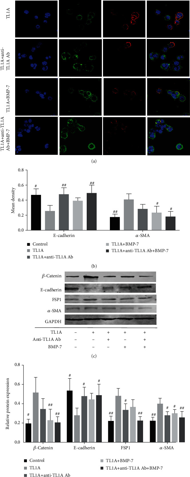
Figure 7.

Inhibition of TL1A in intestinal epithelial cells in vitro using anti-TL1A antibodies and BMP-7. HT-29 cells were exposed to TL1A, anti-TL1A antibodies, and/or BMP-7, respectively. (a, b) The cells were probed with fluorescently labeled antibodies against E-cadherin (red) and FSP1 (green); E-cadherin and FSP1 colocalization (yellow) (1000x) and the mean density of E-cadherin and FSP1 were detected by IPP software. (c, d) Western blot analysis of β-catenin, E-cadherin, α-SMA, and FSP1 in different groups of HT-29 cells. ∗P < 0.05, ∗∗P < 0.01, and ∗∗∗P < 0.001 versus TL1A-treated cells. Data were given as mean ± standard deviation (SD). #P < 0.05, ##P < 0.01, and ###P < 0.001 versus TL1A-treated cells.
