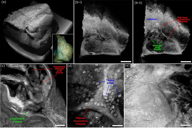
Figure 7.
OCT imaging of unprocessed excised Rattus mammary tumor tissue. (a) Shows a large field OCT image capturing the surface morphology of an entire excised tumor specimen. The inset in the lower right shows a camera image of the same section of tissues. (b) Vertical sections showing subsurface tissue morphology of the tissue captured in (a). Scale Bar: 1.25 mm. (c, d) Small field maximum projection images of different regions of excised Rattus mammary tissues captured using the OCT system. (c) Scale Bar: 100 µm. (d) Scale Bar: 100 µm. (e) Scale Bar: 100 µm.

