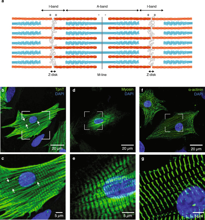
Fig. 1. Myofibrillar organization in neonatal rat cardiomyocytes at the microscale.
a Schematic representation of adjoining micrometer-sized sarcomeres within a myofibril, showing the organization of the thick (cyan) and thin (red) filaments in this basic contractile unit of striated muscle. Neonatal rat cardiomyocytes were labeled with antibodies specific for troponin T (TpnT; b, c), the heavy chain of myosin (Myosin; d, e), and α-actinin (f, g), respectively. Thin (b, c) and thick (d, e) filaments assemble into myofibrils, which align along the main axes of the star-like shaped cells and display regularly spaced Z-disks (f, g). Myofibril branching is indicated with white arrows. c, e, g Zoomed-in views of the framed regions in b, d, f, respectively. A minimum of three biologically independent experiments were performed in each case.

