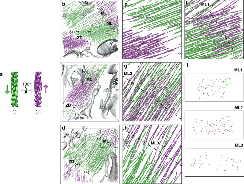
Fig. 4. Structural signature of sarcomere contraction.
a De novo structures of the actin-Tpm filament in opposite orientations, represented by arrows pointing toward the pointed (−) end. b−d Polarity assignment for the neonatal cardiac thin filaments shown in Fig. 2e, f and Supplementary Movie 2 (b; Supplementary Movie 3), Supplementary Fig. 3e, h (c; first half of Supplementary Movie 4) and Supplementary Fig. 3f, i (d; second half of Supplementary Movie 4). Filaments are represented by arrows colored according to the assigned polarity. The gaps between thin filaments facing each other with their barbed ends indicate the location of Z-disks (ZD). The overlap regions between thin filaments of opposite polarity at the pointed (−) ends correspond to M-lines (ML). e, f Zoomed-in views of the framed regions in b showing the thin filament orientations at the Z-disk (e) and M-line (f, ML1). g, h Zoomed-in views of the framed area in c, d, respectively, showing the thin filament orientations at the M-lines (ML2−3). i Cross-sections through ML1−3 at the locations indicated by the dotted lines in f−h. See Supplementary Fig. 8 for visualization of the unassigned filaments.

