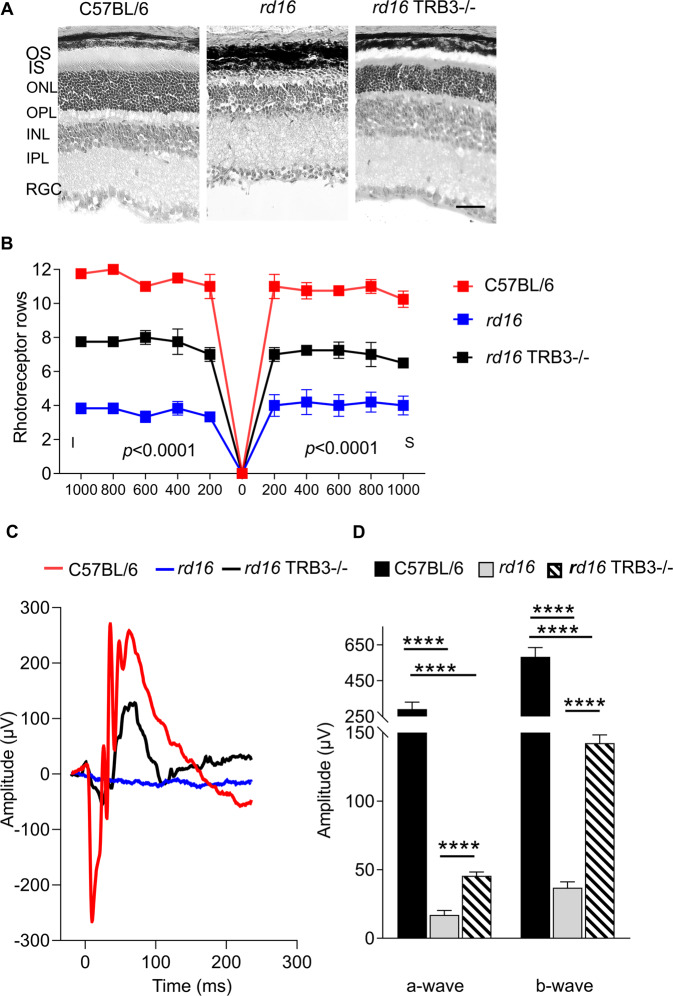
Fig. 3. TRB3 knockout decreases the photoreceptor cell loss detected in P18 rd16 retinas.
A Images of C57BL/6, rd16, and rd16 TRB3−/− retinas stained with eosin and hematoxylin are shown. The scale is 50 µm. B The retinal images were used to calculate the photoreceptor nuclei across the retinas. Preservation of photoreceptor cells was detected in rd16 TRB3−/− mice compared to rd16 mice (n = 4–5). This preservation was in accordance with the improvement of a- and b-scotopic ERG amplitudes registered at P18 (n = 3–7). C Images of the scotopic ERG traces. D ERG analysis demonstrated a statistically significant increase in the a- and b-wave amplitudes in rd16 TRB3−/− retinas. Data are shown as mean ± SEM; ****p < 0.0001. OS outer segments of photoreceptors, IS inner segments of photoreceptors, ONL outer nuclear layer, OPL outer plexiform layer, INL inner nuclear layer, IPL inner plexiform layer, RGC layer retinal ganglion cell layer.

