Fig. 5.
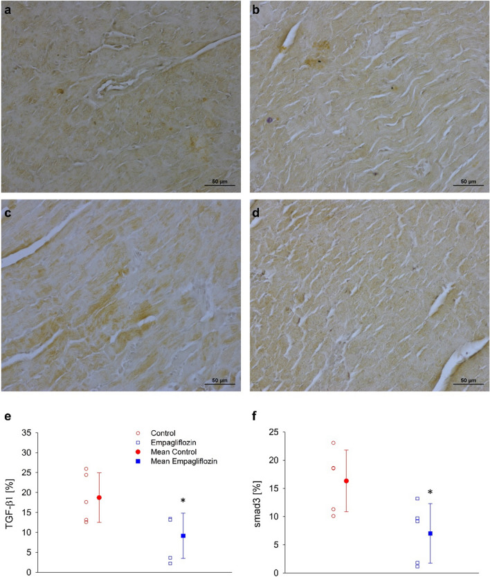
TGF-β1 and Smad3 immuno-histological staining. a Representative photograph of immunoprecipitation of anti-TGF-β1 antibody in control cardiac muscle specimen. b Representative photograph of immunoprecipitation of anti-TGF- β1 antibody in empagliflozin treated rat's cardiac muscle specimen. c Representative photograph of immunoprecipitation of anti-Smad3 antibody in control cardiac muscle specimen. d Representative photograph of immunoprecipitation of anti-Smad3 antibody in empagliflozin treated rat's cardiac muscle specimen. e Expression of TGF-β1. f. Expression of Smad3. Individual measurements (empty symbols) and average (filled symbols, n=6 and 8 to the control and empagliflozin, respectively), control (red) and empagliflozin (blue). * P < 0.05 vs. control, by student's t-test. Photographs magnification x 200.

