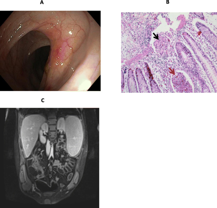
Fig. 1.
Bowel morphology at baseline. A Ileocolonoscopy: ulcerated and ileocecal valve stricture with impossibility to pass through with the scope (Paris classification A1b, L1, B2, G0; SES-CD: 3). B Histology (colonic mucosa): architectural irregularity and a mild patchy increase of lamina propria cells with neutrophilic and eosinophilic infiltration, crypt abscesses (red arrow) and an epithelioid cell granuloma (black arrow) indicating active disease. C Abdomen MRI: active disease with increased wall thickness (max: 10 mm), diffusion restriction and contrast enhancement in the distal ileum (total length:15–20 cm) and ileal stricture; mesenteric hypertrophy (creeping fat) and lymphadenopathy and conglomerated bowel loops (right lower quadrant) are also shown. SES-CD: simplified endoscopic score for Crohn’s disease

