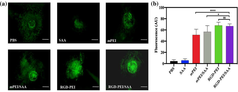
Fig. 8.
Target CNV ability of RGD-PEI/SAA and mPEI/SAA in mice. RGD-PEI/SAA and mPEI/SAA were labeled with FITC and injected (20 µM, 2 µL/eye) into vitreous space immediately after laser-induced CNV. a The RPE/choroid/sclera flat mounts were prepared 72 h after laser injury. The fluorescence in the representative photographs indicates the localization of FITC-labeled RGD-PEI/SAA and mPEI/SAA in the CNV lesions. The white bar represents 100 µm. b The quantitative analysis of RPE/choroid/sclera flat mounts CNV lesions (∗p < 0.05, ****p < 0.0001, ns: no significance)

