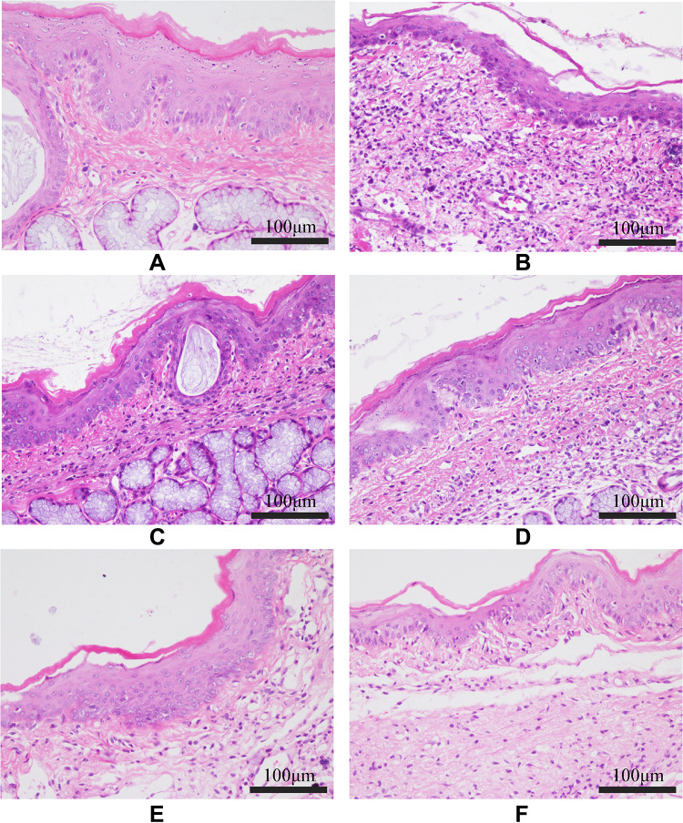
Figure 9.
Histopathological examinations of the pharyngeal mucosal tissue of the rats from each group (HE, 200×). (A) Control group; (B) Model group; (C) FFZJF-L group; (D) FFZJF-M group; (E) FFZJF-H group; (F) AS group. It can be seen that the morphology of the pharyngeal tissues in the control group remain intact based on microscopy. In the model group, the mucous membrane appears necrotic, the submucosal layer shows swelling (edema), and many inflammatory cells are present. The inflammation is significantly reduced in the FFZJF treatment groups. As the dose of FFZJF increases, the condition of inflammation improved.

