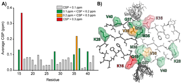
Figure 6:
(A) 13C chemical shift perturbations (CSP) between AβM01-42 and Aβ1-42 plotted as a function of residue position. The only residues where the shifts differ significantly are K16 and V36. (B) Structure of AβM01-42 illustrating the positions of K16 and V36 and other residues with 0.1<CSP<0.2.

