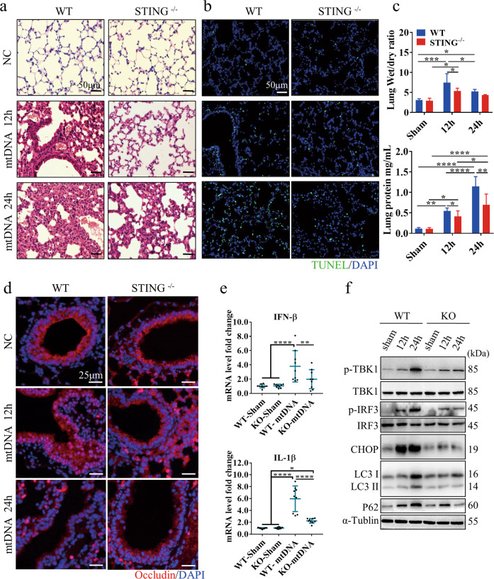
Fig. 2. Circulating mtDNA contributes to ALI by activating the STING pathway and impairing autophagy.
Representative images of lung H&E (a) and TUNEL staining (b) in WT and STING -/- mice at 12 or 24 h after mtDNA administration. The scale bar represents 50 μm. c The lung wet/dry ratio and BALF protein levels in WT and STING-/- mice. d Immunofluorescence staining of occludin in the lung tissue of mice. Red, occludin immunostaining; blue, DNA stained with DAPI. The scale bar represents 25 μm. e qPCR analysis of IFN-β and IL-1β mRNA in the indicated tissues of mice at 24 h after mtDNA injection. f Western blot analysis of STING and autophagy signaling in the lungs of mice at 12 h and 24 h after intraperitoneal injection of mtDNA. Each panel shown represents the mean ± SD taken from at least three independent experiments. *p < 0.05; **p < 0.005; ***p < 0.0001. ns not significant. Two-tailed Student’s t-test was used to determine statistical significance.

