Figure 1.
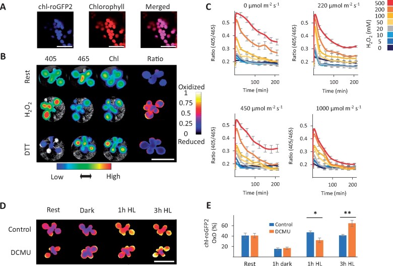
Light-dependent redox modification of chloroplast-targeted roGFP2. A, Confocal images of chl-roGFP2 Arabidopsis plants. Bars = 20 µm. B, Ratiometric analysis of chl-roGFP2 during rest, under oxidized (500 mM H2O2) and reduced (100 mM DTT) conditions using whole-plant imaging. The plants were excited with 405 nm and 465 nm LEDs light sources and fluorescence emission was collected using a 510-nm emission filter. For chlorophyll detection, a 405-nm LED light source and a 670-nm emission filter were used. Color-coded keys represent fluorescence intensities (below image) or 405/465 ratio values (to right of image). Bars = 2 cm. C, The effect of light intensities on chl-roGFP2 reduction after application of H2O2 as examined by whole-plant imaging. Values represent means of six plants ± SE. D and E, The effect of DCMU on chl-roGFP2 oxidation using whole-plant imaging and plate-reader approaches. Oxidation of chl-roGFP2 was detected using whole-plant imaging. Bar = 2 cm. (D) or a plate reader (E). Values for the degree of chl-roGFP2 oxidation (OxD) are presented as means of seven to eight plants ± SE. Asterisks (*) mark significant differences between the ratio of the control/DCMU treatment (t test, P < 0.05, Supplemental File S1). The chl-roGFP2 data were acquired using the chl-TKTP-roGFP2 line.

