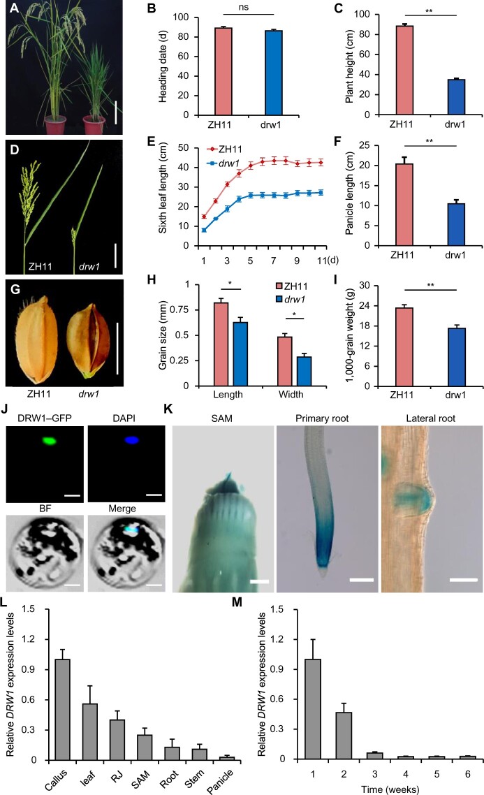
Figure 2.
Mutation of DRW1 causes developmental defects and DRW1 is specifically expressed in dividing cells in rice. (A) Phenotypic performance of wild-type ZH11 and drw1 plants. Bar represents 10 cm. (B–C) Bar graphs of heading date (B) and plant height (C). (D–F) Panicle and leaf morphology. Bar represents 10 cm. (G–I) Kernel morphology. Bar represents 10 mm. (J) Subcellular localization of a DRW1-GFP fusion protein in ZH11 rice protoplasts. Blue fluorescence from DAPI (4,6-diamidino-2-phenylindole) staining indicates the nucleus. Bar = 50 μm. (K) Histochemical GUS assays for stably transformed promoter DRW1-GUS fusion lines. GUS signals appear in the shoot apical meristem and in the meristems of primary and lateral roots. Scale bars: SAM, 1 mm; primary root and lateral root, 100 μm. (L–M) RT-qPCR-based analysis of DRW1 transcription in different tissues (L) and in whole plants from 1 week to 6 weeks of age (M). RJ, rhizome junction. Note that the growth stage of panicle tissue was old (it was 21 cm long). Data are means ± SD (standard deviation), n > 15 in (B–C), (E–F), and (H–I), and n = 3 in (L) and (M). Student’s two-tailed t test (*, p < 0.05; **, p < 0.01; n.s., not significant).

