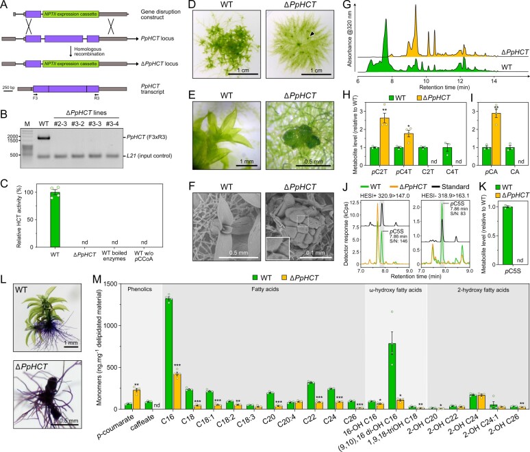
Figure 5.
Investigation of PpHCT function in planta. A, Homologous recombination-mediated strategy for PpHCT gene disruption. A genomic fragment encompassing the second and third PpHCT exons was excised with simultaneous insertion of the NPTII selection cassette conferring resistance to G418. Binding sites of oligonucleotides used for characterization of the transgenic lines are shown. Gray box, UTR; black line, intron; purple box, exon. B, Agarose gel photograph produced from RT-PCR analysis reporting the absence of PpHCT transcripts in the four ΔPpHCT KO lines. M, DNA size marker. C, HCT activity in protein extracts from wild-type (WT) and ΔPpHCT gametophores. HCT activity was measured in vitro using shikimate and p-coumaroyl-CoA as substrates. Negative WT control assays involved boiled protein extracts or omission of p-coumaroyl-CoA (pCCoA). Results are the mean ± SEM of four independent enzyme assays, performed with protein extracts from each of the four independent ΔPpHCT mutant lines. nd, not detected. D, Phenotype of 4-week-old P. patens WT and ΔPpHCT colonies. Arrowhead points to a ΔPpHCT gametophore. E, Magnified image of gametophores visible in (D). F, SEM micrographs of 4-week-old gametophores. For ΔPpHCT, inset shows intercellular adhesive structures (enlargement of boxed region). G, Representative HPLC-UV chromatograms of WT and ΔPpHCT gametophore extracts. H, Relative levels of phenolic threonate esters in gametophore extracts. pC2T, p-coumaroyl-2-O-threonate; pC4T, p-coumaroyl-4-O-threonate; C2T, caffeoyl-2-O-threonate; C4T, caffeoyl-4-O-threonate. I, Relative levels of free hydroxycinnamic acids in gametophore extracts after acid hydrolysis. pCA, p-coumaric acid; CA, caffeic acid. J, Overlay of representative UHPLC-MS/MS chromatograms showing the absence of p-coumaroyl-5-O-shikimate (pC5S) in ΔPpHCT gametophore extracts. Gray shaded regions highlight relevant elution time windows. K, Relative levels of p-coumaroyl-5-O-shikimate (pC5S) in gametophore extracts. Results are the mean ± SEM of three independent WT biological replicates and four independent ΔPpHCT mutant lines. L, Toluidine blue staining of WT and a ΔPpHCT mutant. Protonema and rhizoids do not have a cuticle, and so are readily stained. M, Compositional analysis of WT and ΔPpHCT gametophore cuticular biopolymers. Data are the mean ± SEM of four WT biological replicates and four independent ΔPpHCT mutant lines. WT versus mutant t test adjusted P-value: *P<0.05; **P<0.01; ***P<0.001.

