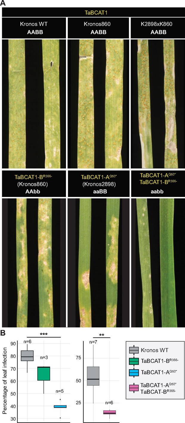
Figure 4.

TaBCAT1 disruption mutants display reduced susceptibility to Pst. A, TaBCAT1-AQ50*, TaBCAT1-BR366-, and TaBCAT1-AQ50* TaBCAT1-BR366- disruption mutants all displayed limited sporulation, higher degrees of necrosis and less chlorosis when infected with Pst isolate 13/14 and compared with the Kronos wild-type (WT). Negative controls included Kronos WT and Kronos ethyl methanesulfonate mutants (Kronos860 and K2898xK860) carrying WT alleles of TaBCAT1. Images were captured 20 dpi. B, The percentage of leaf infection was significantly reduced in TaBCAT1 disruption mutants at 20 dpi. Asterisks denote statistically significant differences between each pair of conditions (***p < 0.001, **p < 0.01, two-tailed t-test). Bars represent median values, boxes signify the upper (Q3) and lower (Q1) quartiles, and whiskers are located at 1.5 the interquartile range
