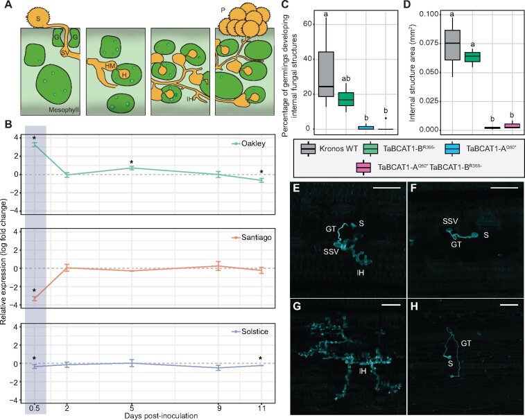
Figure 5.
TaBCAT1 expression early during Pst infection is required for susceptibility. A, A controlled time-course of infection was carried out with Pst isolate F22 and wheat varieties Oakley, Solstice and Santiago. S, urediniospore; SV, sub-stomatal vesicle; IH, invasive hyphae; HM, haustorial mother cell; H, haustorium; P, pustule; G, guard cell. B, During Pst infection, TaBCAT1 expression at 12 hpi was highest in the most susceptible variety Oakley, whereas the most resistant variety Santiago displayed a significant reduction in TaBCAT1 expression (highlighted area). Two independent leaves from the same plant were pooled and three independent plants analyzed for TaBCAT1 expression by RT-qPCR at 12 hpi, 2 dpi, 5 dpi, 9 dpi, and 11 dpi. TaBCAT1 expression was compared between Pst-infected and mock-inoculated plants for each time point per variety. Asterisks denote statistically significant differences (***p < 0.005, **p < 0.01, *p < 0.05; two-tailed t test). Error bars represent standard deviations. C, D, Histological studies using a fungus-specific fluorescent dye revealed differences in the extension of internal fungal structures between WT and the TaBCAT1-AQ50* TaBCAT1-BR366- disruption mutant. The number of germinating spores assessed was as follows: Kronos WT n = 229, TaBCAT1-AQ50*n = 114, TaBCAT1-BR366-n = 297, TaBCAT1-AQ50* TaBCAT1-BR366-n = 152. The number of internal structures measured was as follows: Kronos WT n = 9, TaBCAT1-AQ50*n = 2, TaBCAT1-BR366-n = 10, TaBCAT1-AQ50* TaBCAT1-BR366-n = 3. Letters indicate significant differences determined using Duncan’s multi range test (p < 0.05). Bars represent median values, boxes signify the upper (Q3) and lower (Q1) quartiles, and whiskers are located at 1.5 the interquartile range. E, WT plants showed clear Pst hyphal development at 4 dpi. F, Hyphal growth was not observed for TaBCAT1-AQ50* TaBCAT1-BR366- at 4 dpi. G, At 6 dpi WT plants showed large, intricate fungal structures. H. The TaBCAT1-AQ50* TaBCAT1-BR366- disruption mutant at 6 dpi showed a unique phenotype where germinating spores reached stomata and entered the underlying sub-stomatal space, but further fungal growth was absent. Scale bars in E–H represent 100 µm; S, urediniospore; GT, germ tube; SSV, sub-stomatal vesicle; IH, invasive hyphae

