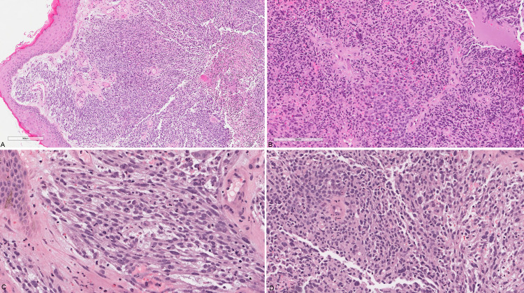
Figure 2. Valvular biopsy.
(A) Low power view showing unremarkable squamous epithelium overlying sheets of neoplastic cells; (B) diffuse sheets of pleomorphic plump to spindle neoplastic cells; (C) and (D) high power view of the spindle cell areas showing brisk mitosis and focal possible rhabdomyoblastic differentiation.

