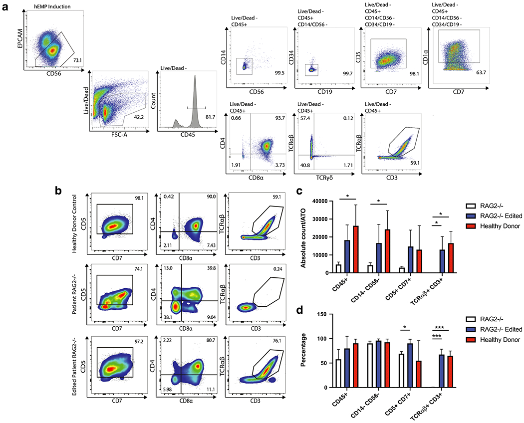
Figure 2: Human iPSC derived T cell differentiation in healthy donor cells and in RAG2 deficient and RAG2 edited cells.

A. Representative analysis of the flow cytometry gating showing expression of early and mature T cell differentiation markers for a healthy donor sample. B. Flow cytometry results of T cell differentiation markers for the healthy donor cells, the RAG2 deficient patient cells, and the RAG2 gene-edited cells showing co-expression of CD5 and CD7, along with CD4, CD8α, and in the case of the healthy donor and edited cells, also CD3 and TCRαβ. C. Absolute count of indicated iPSC-derived T cell subsets per ATO in the healthy donor sample, the RAG2 deficient sample, and the gene-edited sample. D. Representative data showing differentiation profiles for the three examined samples over three distinct experiments. Statistical significance was computed using multiple -tests with 1% False Discovery Rate with two-stage step-up method of Benjamini, Krieger and Yekutieli. *, p<0.05; ***, p<0.001
