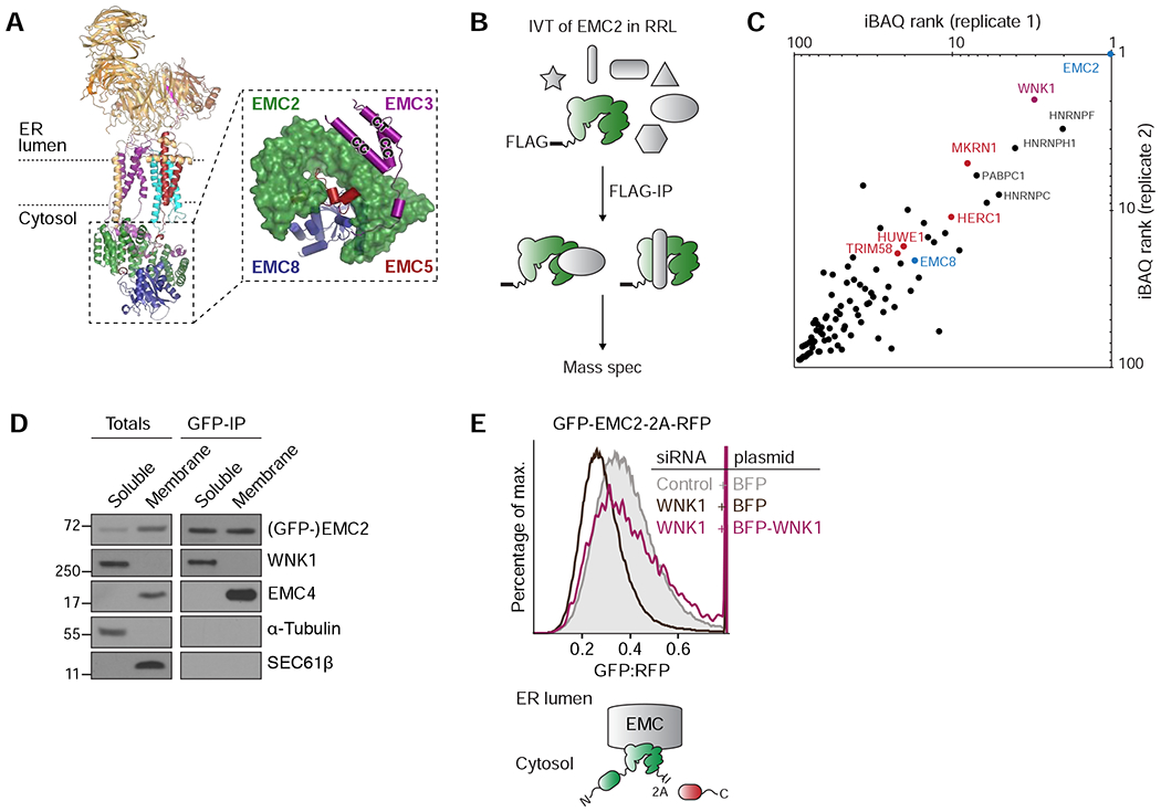
Figure 1. Unassembled EMC2 binds WNK1.

(A) Overview of the cytosolic domain of the human EMC, assembled around EMC2 (PDB 6WW7; (Pleiner et al., 2020). Inset: close-up on the cytosolic domain as viewed from the membrane. CC = coiled coil; CT = C-terminus. (B) Strategy for identification of binding partners of unassembled EMC2. 3xFLAG-tagged EMC2 was in vitro translated (IVT) in rabbit reticulocyte lysate (RRL) and isolated by native immunoprecipitation (IP) with anti-FLAG affinity resin and analyzed by mass spectrometry. (C) EMC2-specific interaction partners, identified by mass spectrometry as outlined in B, with an LFQ-ratio > 5 over controls were ranked by their iBAQ values (see Table S2). The resulting top hits from two replicates are plotted. (D) Cells stably expressing GFP-EMC2 were fractionated into cytosolic and membrane-associated fractions. GFP-tagged EMC2 was purified from both fractions under native conditions using an anti-GFP nanobody, and its interaction partners were analyzed by Western blotting with the indicated antibodies. (E) HEK293T cells stably expressing GFP-EMC2 were transiently transfected with plasmids encoding either BFP or BFP-tagged siRNA-resistant WNK1. Cells were treated with either a scrambled control or WNK1 siRNA, and BFP-positive cells were analyzed by flow cytometry. The relative levels of GFP-EMC2, normalized to an internal RFP expression control (described in methods), are plotted as a histogram and reflect changes in subunit stability.
