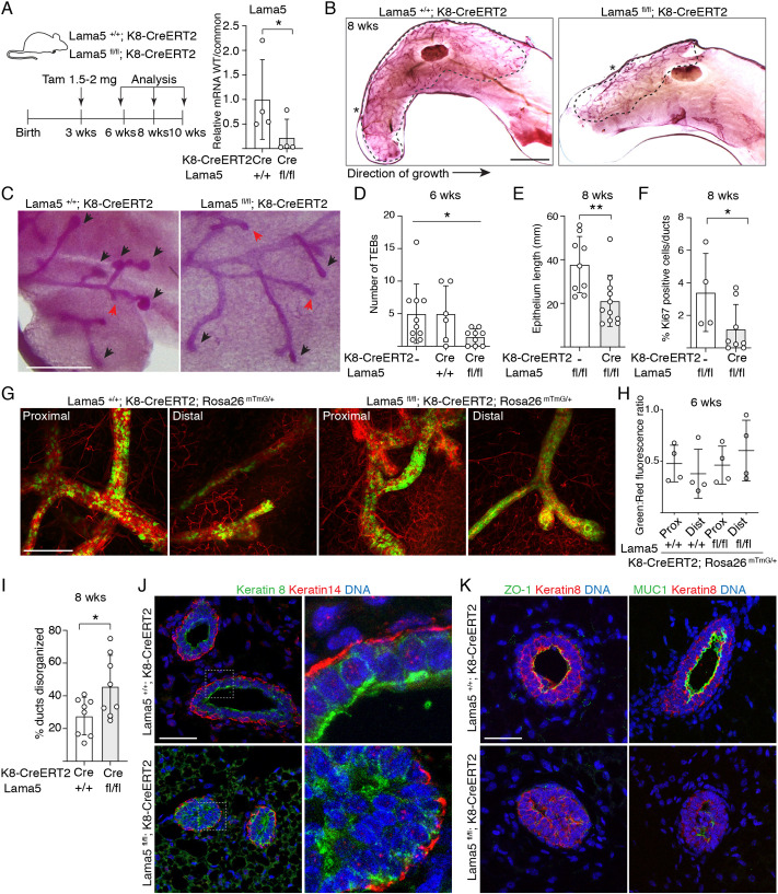
Fig. 2.
Luminal laminin α5 is required for pubertal growth of the mammary epithelium. (A) Schematic showing the outline of tamoxifen induction of K8-CreERT2 during puberty, and qPCR analysis of Lama5 mRNA expression in luminal MECs (+/+ and fl/fl, n=4 samples analyzed) by using wild-type (WT) allele-specific primers compared with primers common for both + and flox alleles (common). (B) Representative images of carmine alum-stained #4 mammary glands of 8-week-old Lama5+/+;K8-CreERT2 and Lamafl/fl;K8-CreERT2 treated with tamoxifen. The dashed line shows the outline of ductal growth and the asterisks mark the beginning of epithelium. (C) Representative images of TEBs and ductal ends (marked by black arrowheads and red arrows, respectively) in 6-week-old transgenic mice. (D) Quantification of the number of TEBs in 6-week-old transgenic mice analyzed from #4 mammary glands (K8-CreERT2, n=10; Lama5+/+;K8-CreERT2, n=6, Lama5fl/fl;K8-CreERT2, n=9 glands analyzed). (E) Quantification of epithelium length in 8-week-old transgenic mice analyzed from #4 mammary glands (Lama5fl/fl; n=9, Lama5fl/fl;K8-CreERT2, n=11 glands analyzed). (F) Quantification of Ki67 positivity in mammary glands of 8-week-old transgenic mice (Lama5fl/fl; n=4; Lama5fl/fl;K8-CreERT2, n=8 individuals analyzed). (G) Representative images of #4 mammary glands of 6-week-old Lama5+/+;K8-CreERT2;R26mTmG/+ and Lama5fl/fl;K8-CreERT2;R26mTmG/+ mice. Images show proximal (closer to the beginning of epithelium) and distal parts of the duct. (H) Quantification of green to red fluorescence ratio in proximal and distal parts of the duct in Lama5+/+;K8-CreERT2;R26mTmG/+ and Lama5fl/fl;K8-CreERT2;R26mTmG/+ mice (+/+ and fl/fl, both proximal and distal part, n=4 glands quantified). (I) Quantification of epithelial disorganization in ducts of 8-week-old Lama5+/+;K8-CreERT2 (n=8) and Lama5fl/fl;K8-CreERT2 (n=8) animals. (J) Representative immunofluorescence images of 8-week-old Lama5+/+;K8-CreERT2 and Lama5fl/fl;K8-CreERT2 glands immunostained with K8 and K14 antibodies. Boxes indicate the magnified regions in the right panels. (K) Representative immunofluorescence images of 8-week-old Lama5+/+;K8-CreERT2 and Lama5fl/fl;K8-CreERT2 glands immunostained with ZO-1 or MUC1, and K8 antibodies. Data points indicate analysis of individual mice. Data are mean±s.d. *P<0.05, **P<0.01 [unpaired two-tailed Student's t-test (A,E,I) or Welch's ANOVA test (D)]. Scale bars: 5 mm (B); 0.5 mm (C); 200 µm (G); 50 µm (J,K).

