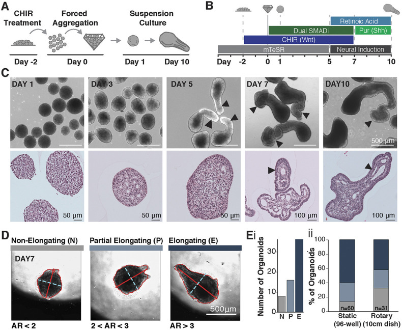
Fig. 1.
CHIR treatment of neural PSC aggregates results in axial extension. (A,B) Schematic of experimental set-up and differentiation protocol using 2 μM CHIR. (C) Bright-field and histological images of elongating organoids (arrowheads indicate elongated structures). (D) Images from time-course of extension, displaying organoids that do not elongate (N), partially elongate (P) or fully elongate (E) with respective axis ratios (ARs) of AR<2, 2<AR<3 and AR>3 (minor axis dotted cyan line, major axis solid red line). (E,i) Elongation types detected during time lapse imaging (n=60 organoids). (E,ii) Quantification of elongating types across static (n=60 organoids) and rotary (n=31 organoids) cultures.

