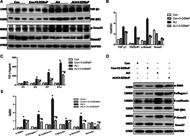
Fig. 6.
3-DZNeP lessens the in vivo and in vitro epithelial to mesenchymal transition (EMT) by inhibiting activation of TGF-β/Smad signaling pathways. A Lung tissue lysates on Day 14 after intratracheal LPS or PBS in the control, control + 3-DZNeP, ALI, ALI + 3-DZNeP groupswere subjected to immunoblot analysis with specific antibodies against TGF-β1, TGF-βR1, p-Smad2, Smad7 and GAPDH. B Expression levels of TGF-β1, TGF-βR1, p-Smad2 and Smad7were quantified by densitometry and normalized using GAPDH. C BALF was harvested on Day 1, Day 3, Day 7 and Day14 after intratracheal LPS in the control, control + 3-DZNeP, ALI, ALI + 3-DZNeP groups. The expression levels of TGF-β1 were measured by ELISA. All data are expressed as mean ± SEM. (n = 9–15/group, * P < 0.05 vs. control or 3-DZNeP group, #P < 0.05 vs. ALI group, determined by one-way ANOVA for multiple group comparisons). D, E Alveolar macrophages isolating from different treatment ALI mice on Day 14 after intratracheal LPS or PBS were co-cultured with mouse lung epithelial cell lines (MLE-12). Cell lysates were subjected to immunoblot analysis with specific antibodies against α-SMA, Collagen I, E-cadherin, TGF-βR1, p-Smad2, Smad7 and GAPDH (D).Expression levels of α-SMA, Collagen I, E-cadherin, TGF-βR1, p-Smad2, Smad7 were quantified by densitometry and normalized using GAPDH. All data are expressed as mean ± SEM. (n = 4–6/group, *P < 0.05 vs. control or 3-DZNeP group, #P < 0.05 vs. ALI group, determined by one-way ANOVA for multiple group comparisons)

