
FIGURE 1.
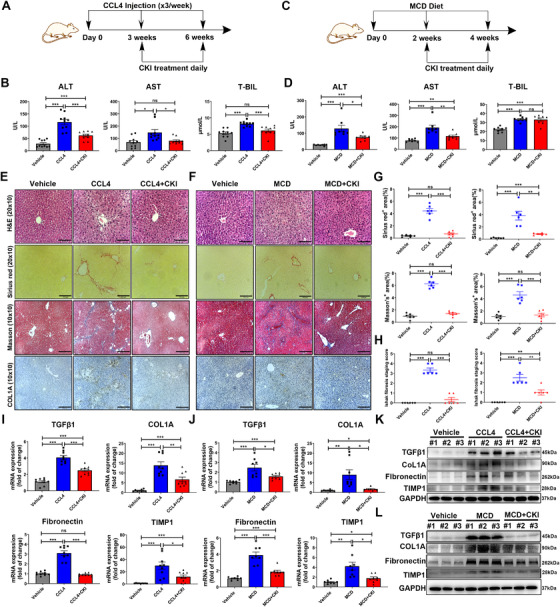
CKI attenuates chronic liver fibrosis. (A) Scheme of experimental procedure for C57BL/6 mice intraperitoneally treated with 4 ml/kg CCl4 in olive oil for 6 weeks. Mice were intraperitoneally administrated with CKI (7.5 ml/kg) for 3 weeks, starting at 3 weeks post initiation of CCl4 challenge. (B) Serum levels of ALT, AST, and T‐BIL were detected after the final CKI treatment in CCl4‐treated mice (n = 10). (C) Scheme of experimental procedure for C57BL/6 mice fed with MCD diet for 4 weeks. Mice were intraperitoneally administrated with CKI (7.5 ml/kg) for 2 weeks, starting at 2 weeks post initiation of MCD diet challenge. (D) Serum levels of ALT, AST, and T‐BIL were quantified after the final CKI treatment in MCD diet‐treated mice (n = 8). (E and F) Mice liver sections from CCl4‐induced or MCD diet‐induced liver fibrosis models were collected for H&E (original magnification 20 × 10, scale bar 110 μm), Sirius Red (original magnification 20 × 10, scale bar 100 μm), Masson staining (original magnification 10 × 10, scale bar 220 μm), and collagen type1 (COL1A, original magnification 10 × 10, scale bar 210 μm) immunostaining after the final CKI treatment (n = 6). (G) Positive Sirius Red (above) or Masson staining (below) area were quantified by ImageJ analysis (n = 6). (H) Ishak fibrosis score of the Sirius Red‐stained liver sections (n = 6). (I and J) mRNA expression of TGF‐β1, COL1A, Fibronectin, and TIMP1 were analyzed by qRT‐PCR in liver tissues from CCl4‐challenged (n = 9) or MCD diet‐challenged (n = 8) mice. (K and L) Western blot assay for detecting the expression of TGF‐β1, COL1A, Fibronectin, and TIMP1 in liver tissues from CCl4‐challenged (K) or MCD diet‐challenged (L) mice. Data are presented as means ± SEM. ns, p > 0.05; *p < 0.05; **p < 0.01; ***p < 0.001