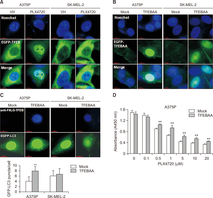
Fig. 3.
TFEB nuclear translocation and autophagy induction in PLX4720-treated A375P cells. (A) Cells were transiently transfected with pEGFP-TFEB, exposed to PLX4720 (1 μM) for 24 h, and then fixed and counter-stained with Hoechst 33342 to identify the nucleus. (B) Cells were transiently transfected with FLAG-tagged TFEBAA (constitutively active S142A/S211A-TFEB mutant), and then fixed and counter-stained with Hoechst 33342 to identify the nucleus. (C) A375P and SK-MEL-2 cells were co-transfected with FLAG-tagged TFEBAA and pEGFP-LC3 before being stained with anti-FLAG antibodies. TFEBAA localization and LC3 puncta formation were detected by fluorescence microscopy. Lower panel: quantification of GFP-LC3 puncta per cell. Data are expressed as mean ± SD. **p<0.01 as compared with mock-transfected cells, as determined by unpaired t-test. (D) A375P cells were transfected with mock or FLAG-tagged TFEBAA for 24 h. The cells were washed, treated with increasing concentrations of paclitaxel ranged from 0.1 to 0.5 μM for 72 h. Cell viability was determined by the WST-1 assay. All values are relative to the vehicle control and are presented as mean ± SD (n=4). **p<0.01 as determined by the Dunnett’s t-test compared to control cells. Values represent the mean ± SD of quadruplicate determinants from one of three representative experiments.

