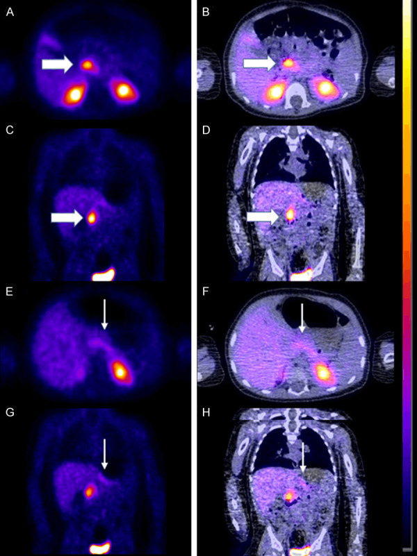
Figure 3.

Preoperative 18F-FDOPA PET scan in a 6-month-old baby boy demonstrating the focal form of CHI. A. Axial PET. B. Axial fused PET-CT. C. Coronal PET. D. Coronal fused PET-CT showing intense uptake in the head of the pancreas (thick white arrow). E. Axial PET. F. Axial fused PET-CT. G. Coronal PET. H. Coronal fused PET-CT showing only very mild uptake in the tail of the pancreas (thin white arrow).
