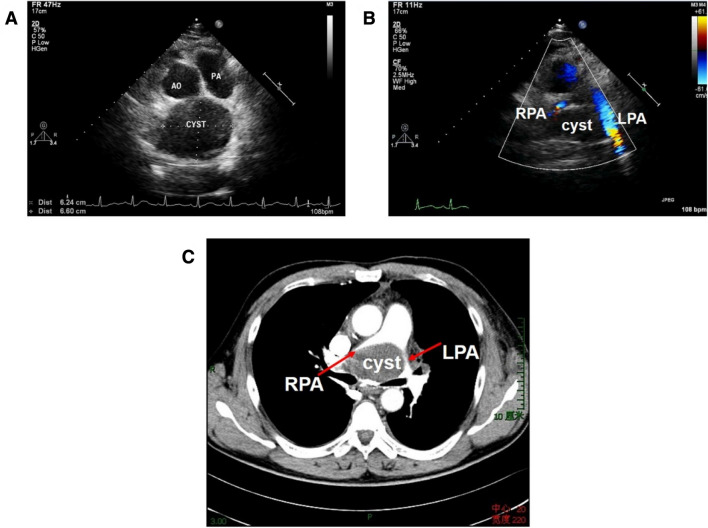
Fig. 3.
A 47-year-old man with a bronchogenic cyst, complaining of dyspnea and chest pain. a Nonstandard parasternal short-axis view of great arteries showed an echo-free structure in the upper region to the heart; b CDFI demonstrated accelerated blood flows in pulmonary branches resulting from the compression on bifurcation of the pulmonary artery; c Contrast-enhanced CT demonstrated the compression of the cyst. PA pulmonary artery, LPA left pulmonary artery, RPA right pulmonary artery

