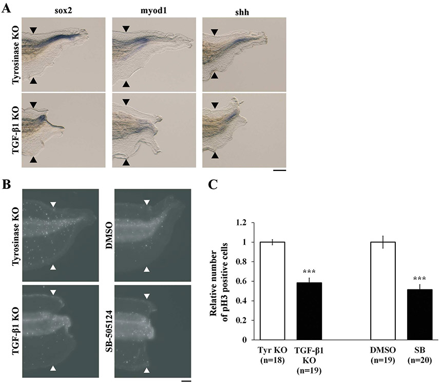
Figure 3. TGF-β1 regulates tissue differentiation and cell proliferation.
(A) Lateral views of WISH performed with sox2 (spinal cord), myod1 (muscle) and shh (notochord) antisense RNA probes at 72 hpa. Scale bar, 200 μm. (B) Whole-mount immunostaining of phosphorylated Histone H3 (pH3) at 48 hpa. Scale bar, 100 μm. (C) Quantification of mitotic cells in the regenerating tail. The number of pH3 positive cells in tgfb1 KO and SB-505124-treated tadpoles was normalized against tyrosinase KO and DMSO-treated control tadpoles, respectively. Black and white arrowheads indicate amputation sites. ***P < 0.001.

