
Figure 4.
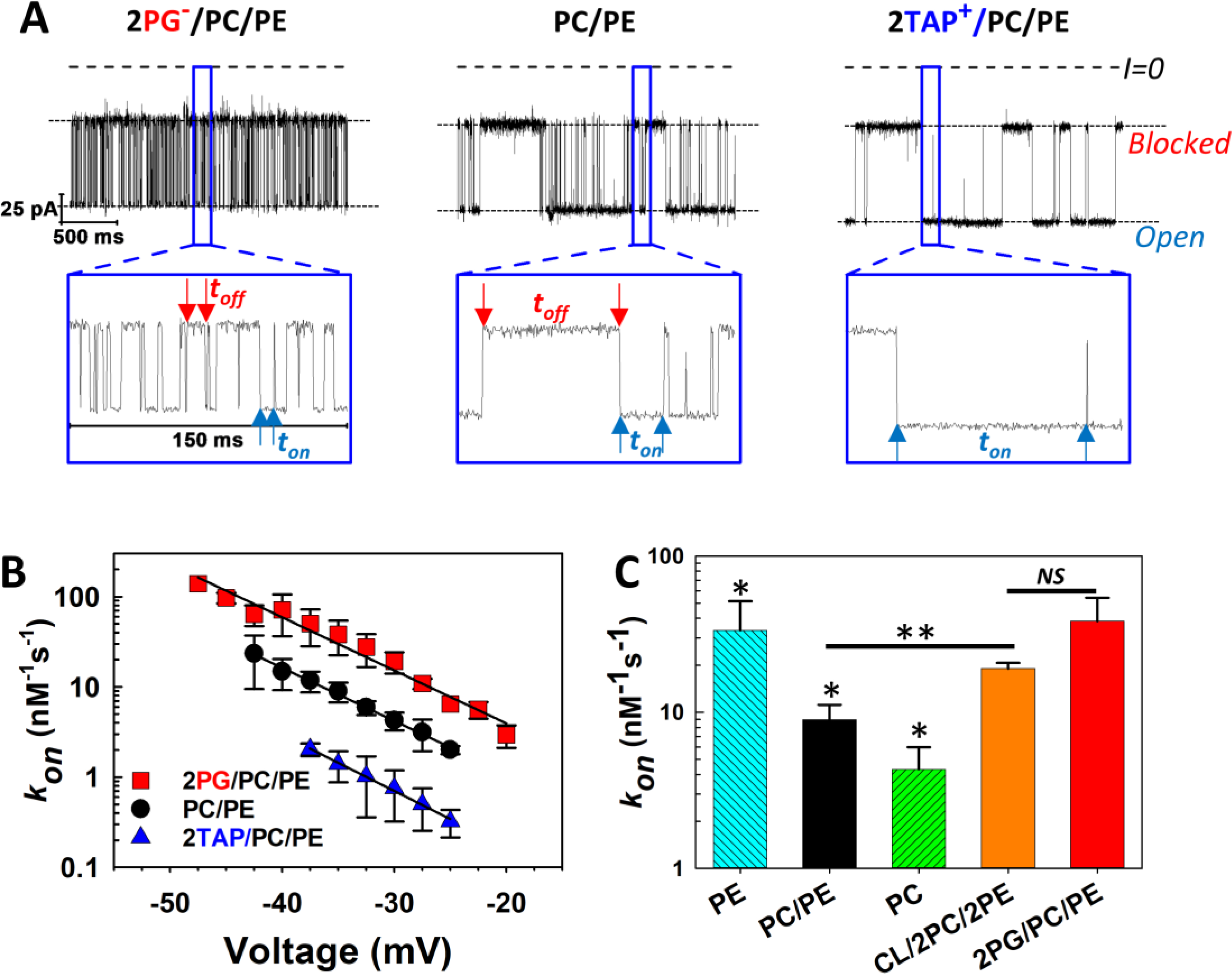
The kinetics of αSyn blockage of VDAC strongly depend on membrane lipid composition. (A) Records of ion currents through single VDAC1 channels reconstituted into planar bilayers formed from DOPG:DOPC:DOPE (2:1:1, mol:mol) (2PG/PC/PE) (left trace), DOPC:DOPE (1:1, mol:mol) (PC/PE) (middle traces), and DOTAP:DOPC:DOPE (2:1:1, mol:mol) (2TAP/PC/PE) (right trace) obtained at −35 mV applied voltage. Individual, time-resolved blockage events can be seen in the insets, which show fragments of current records at a finer time scale. Horizontal dotted lines indicate VDAC open and blocked states; dashed lines indicate zero current. Blue arrows indicate durations of the open state, ton, and red arrows show blocked state durations, toff. Both parameters of blockage events visibly depend on lipid composition. Current traces were digitally filtered using a 5 kHz lowpass Bessel filter for presentation. (B) Voltage dependences of the rate of capture, kon, obtained in three lipid compositions. The kon of the αSyn-VDAC interaction increases in the presence of anionic and nonlamellar lipids. Error bars show 68% confidence intervals. (C) Summary of results for kon obtained in DOPE (PE), PC/PE, DOPC (PC), Cardiolipin:DOPC:DOPE (1:2:2) (CL/2PC/2PE), and 2PG/PC/PE membranes at −35 mV applied voltage. All data were obtained in the presence of 10 nM of αSyn in the cis compartment in 1 M KCl at pH 7.4. Error bars show 68% confidence intervals. Adapted with permission from Jacobs et al. Sci. Reports (2019). Copyright © 2019 Springer Nature.