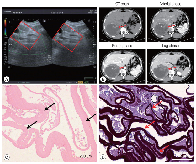
Fig. 1.
Imaging and pathological findings of liver lesions. (A) Color Doppler ultrasonography of the liver. A hybrid mass 11.6×11.2 cm was observed in the right posterior lobe of the liver (red box). (B) Abdominal dynamic phase III computed tomography (red arrow). Round low-density shadows were seen in the right lobe of the liver, and no enhancement was observed after contrast-enhanced scan. High-density nidi were scattered in the lesion. The maximum cross-section of the lesion is about 9.6×8.9 cm. (C) The hematoxylin and eosin stain of paraffin sections displayed the laminated layer (black arrows). (D) Periodic acid-Schiff (PAS) stain presented a strongly PAS-positive basophilic laminated layer (red arrows).

