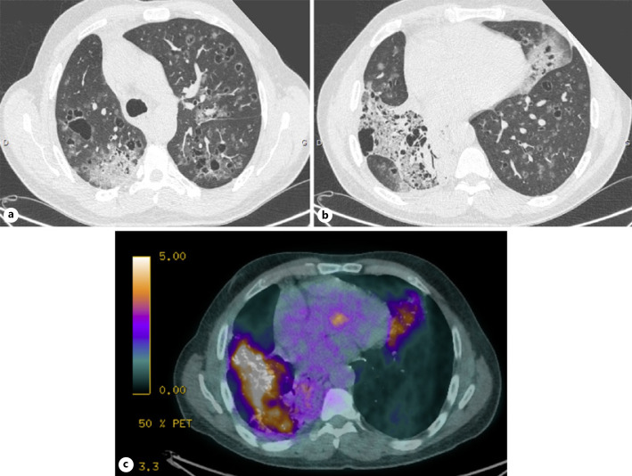
Fig. 1.
CT scan with extensive bilateral pulmonary cystic opacities, surrounded by areas of ground-glass opacity (a); CT scan with extensive bilateral pulmonary cystic opacities, surrounded by areas of ground-glass opacity (b); PET-CT scan with intensely hypermetabolic right lower lobar fibrotic range (c).

