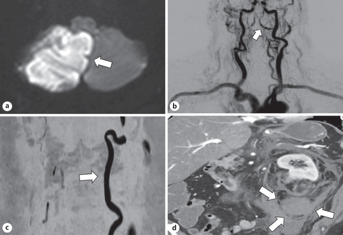
Fig. 1.
a Restricted diffusion in the right posteromedial inferior cerebellum (arrow) compatible with a right PICA subacute infarction. No hematoma is identified. b Early phase of a reconstructed dynamic contrast MRA brain and neck. The left vertebral artery is well visualized throughout its course. There is some reflux of contrast in the right vertebral artery from the left vertebral artery (arrow). c Noncontrast MRA set to display flow in the rostral direction. Near the craniovertebral junction, only flow in the left vertebral artery is seen (arrow). d CT abdomen axial showing a defect in the lower pole of the left kidney containing fat density measuring <4 cm in diameter (AML) with contiguous large retroperitoneal hemorrhage (arrows). PICA, posterior inferior cerebellar artery.

