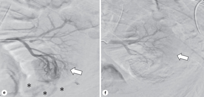
Fig. 2.
e Pre-embolization arterial phase angiogram showing a hypervascular blush (arrow) in the right inferior renal pole with active contrast extravasation extending inferior to the kidney in multiple streams (asterisks). f Post-embolization arterial phase angiogram showing normal vascularization of the upper pole of the left kidney and hypovascularity of the lower pole (arrow) without active contrast extravasation.

