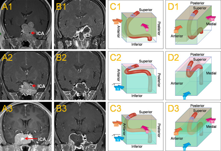
Figure 1.
This shows the common location of the horizontal ICA in Knosp grade 4 PAs and the main surgical approaches. When the horizontal ICA was inferior to the tumor (A1), it caused the horizontal ICA to sink due to the oppression of the tumor, resulting in a larger posterosuperior compartment and a smaller anteroinferior compartment within the CS. The medial approach, often combined with the superior-lateral approach was mainly used to remove the tumor (C1, D1). Postoperative MRI revealed a subtotal resection of the tumor (B1).When the horizontal ICA was superior to the tumor (A2), it caused the horizontal ICA to be raised by the tumor, leading to a smaller posterosuperior compartment and a larger anteroinferior compartment within the CS. The anteroinferior approach was mainly used to remove the tumor (C2, D2). Postoperative MRI revealed a total resection of the tumor (B2). When the horizontal ICA was in the middle of the CS tumor (A3), which was a combination of the two conditions, then the medial, superior-lateral, and anteroinferior approaches could be used (C3, D3). Postoperative MRI revealed a total resection of the tumor (B3). The red, thick arrow ‘M’ represents the medial approach, the orange-yellow, thick arrow ‘SL’ represents the superior-lateral approach, and the blue, thick arrow ‘A’ represents the anteroinferior approach.

