Fig. 5.
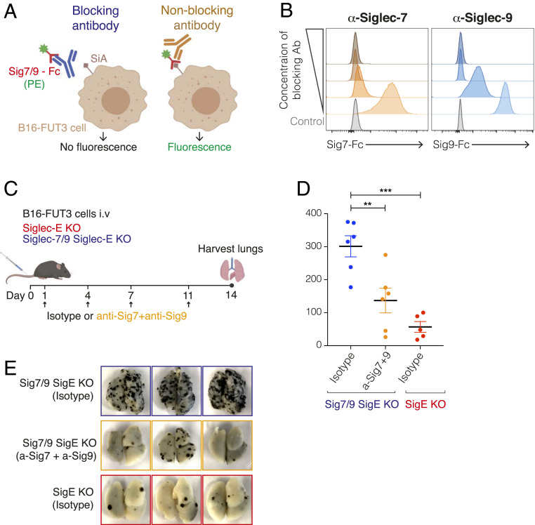
Siglec-7– and Siglec-9–targeting antibodies reduce tumor burden in mice. (A) Illustration of the experimental design to evaluate the blocking ability of different Siglec-7 and Siglec-9 antibody clones. Siglec-7–Fc or Siglec-9–Fc chimeras (hIgG) were preincubated with anti–Siglec-7 or anti–Siglec-9 antibodies (with a mIgG1 D265A Fc), respectively, followed by incubation with B16-FUT3 cells (that express high density of Siglec ligands, SiA). Binding of Siglecs to tumor cells was detected by a fluorescently labeled anti-human IgG antibody that bound to the Fc of the Siglec-Fc chimeras. (B) Binding of Siglec-7–Fc (Left) or Siglec-9–Fc (Right) chimeras to B16-FUT3 cells in the presence of increasing concentrations of anti–Siglec-7 (Left) or anti–Siglec-9 (Right) antibodies. (C) Illustration of experimental design. Mice were inoculated intravenously with B16-FUT3 cells and treated with a combination of anti–Siglec-7 + anti–Siglec-9 antibodies or an isotype-matched control on days 1, 4, 7, and 11 (arrows). On day 14 mice were killed, lungs were excised and fixed, and the number of lung metastatic foci was counted. (D) Number of B16-FUT3 metastatic foci in the lungs of Siglec-7/9/Siglec-E KO mice that received an isotype control (blue), or a combination of anti–Siglec-7 + anti–Siglec-9 antibodies (orange). Siglec-E KO mice that received an isotype control (red) served as control. Each circle represents an individual mouse. Graph shows a representative experiment (n = 2 experiments). Data are displayed as mean ± SEM **P < 0.01; ***P < 0.005 (one-way ANOVA). (E) Representative images of lungs excised from the indicated mouse groups 14 d postinoculation with B16-FUT3 cells.

