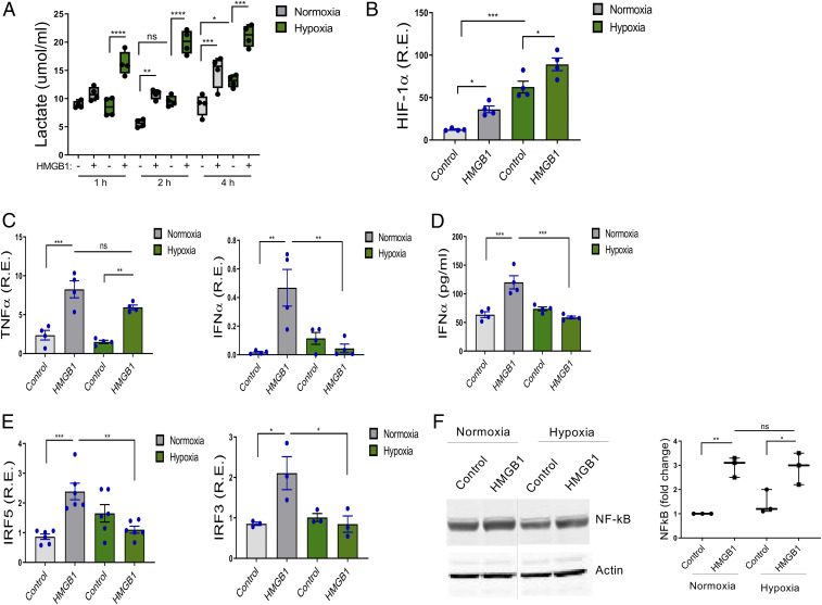
Fig. 1.
Hypoxia reduces HMGB1-induced IRF5, IRF3, and IFNα but not TNFα. (A) Human monocytes were exposed to 21% (normoxia) and 2% (hypoxia) oxygen with or without HMGB1 (1 μg/mL). Lactate measured from cell supernatants at indicated time points demonstrated significantly increased lactate in cells exposed to hypoxia and HMGB1. Floating bars (minimum to maximum), the line at median, each symbol represents an individual experiment in triplicate, n = 4. (B–D) The expression of the HIF-1α, TNFα, IFNα, IRF5, and IRF3 were analyzed by qRT-PCR (4 h). Data represent mean ± SEM of four independent experiments in which each condition was tested in triplicate. (E) The level of IFNα secretion was determined by ELISA from cell supernatant at 24 h. n = 4. (F) NF-κB was induced by HMGB1 in both normoxic and hypoxic cells. NF-κB was analyzed by Western blots in cells under resting and HMGB1-stimulated, normoxic and hypoxic conditions. One representative of three independent experiments (Left). Fold changes compared to normoxic control were calculated by band intensity (Right). The lanes were run on the same gel but were noncontiguous (dotted line). n = 3. Each symbol represents an individual experiment. One-way ANOVA *P ≤ 0.05; **P ≤ 0.01; ***P ≤ 0.001; ns, P > 0.05.

