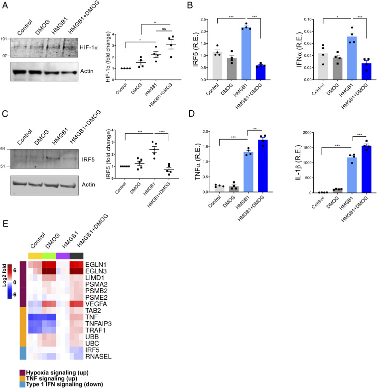
Fig. 2.
DMOG and HMGB1 down-regulate IRF5 and IFN signaling and mimic hypoxia conditions. Human monocytes were preincubated with DMOG (25 μM) for 1 h and stimulated with HMGB1 (1 μg/mL) for 4 h. Each condition was tested in triplicate. (A) Western blots analyzed the level of HIF-1α. One representative of four independent experiments (Left). Fold changes compared to control were calculated by band intensity (Right). (B) IRF5 or IFNα mRNA. Mean ± SEM, n = 4 (C) IRF5 protein. Fold changes compared to untreated control were calculated by band intensity. Mean ± SEM, n = 5. (D) TNFα or IL-1β mRNA. Mean ± SEM, n = 4. One-way ANOVA. *P ≤ 0.05; **P ≤ 0.01; ***P ≤ 0.001; ns, P > 0.05. (E) A heat map generated from RNA-seq of control, DMOG-, HMGB1-, and HMGB1 plus DMOG–treated monocytes (4 h). Two donors were analyzed in triplicate.

