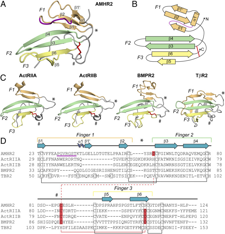
Fig. 4.
Comparison of the TGF-β family type II receptor structures. (A) Structure of AMHR2 ECD with finger 1 (F1, light orange), finger 2 (F2, light green), and finger 3 (F3, light yellow) labeled. Finger 1 loop in AMHR2 is colored in purple. Secondary structure elements are labeled. * indicates the finger 1/2 loop, and # indicates the finger 2/3 loop. The shuffled disulfide bond is highlighted in red sticks. (B) Topology map of AMHR2 with fingers colored and labeled as in A. (C) Structures of ActRIIA (PDB ID 2GOO), ActRIIB (PDB ID 2H3K), BMPR2 (PDB ID 2HLQ), and TβR2 (PDB ID 1M9Z) ECD with fingers and features labeled as in A. (D) Sequence alignment of type II receptors. Fingers 1 to 3 and loops are indicated by the brackets or symbols above the sequence colored and labeled as in A. Secondary structural features are labeled above the sequence accord to the AMHR2 ECD structure. Cysteines are outlined with a black box, and conserved disulfide pairs are indicated by black solid lines. The nonconserved disulfide is highlighted in red. The solid red line shows the disulfide pair in ActRIIA/ActRIIB/BMPR2, while the dotted red line shows the disulfide pair in AMHR2. Residue numbers are labeled at the beginning and end of each line.

