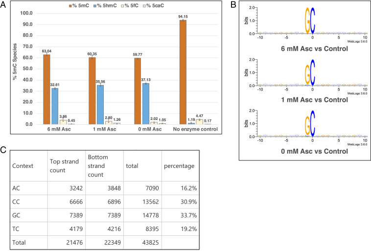
Fig. 6.
(A) LC-MS/MS data showing 5mC oxidation in vitro on Xp12 gDNA (6 ng/μL or 6.3 μM 5mC) by TET43 (20 μM) in 50 mM MES, pH 6.0, 70 mM NaCl, 5 mM 2OG, and 80 μM Fe(II) and varying concentrations of ascorbate (6, 1, or 0 mM). The reactions were incubated for ∼17 h at 37 °C. Error bars represent the SD, n = 3. (B) Sequence logo plots of Xp12 methyl hydroxylation motifs by TET43. Experimental details are found in SI Appendix, SI Materials and Methods. (C) Calculation of percent NC sites in Xp12 DNA. As Gp5mC constitutes 33.7% of all 5mC in Xp12, the concentration of Gp5mC is calculated to be 2.1 μM.

