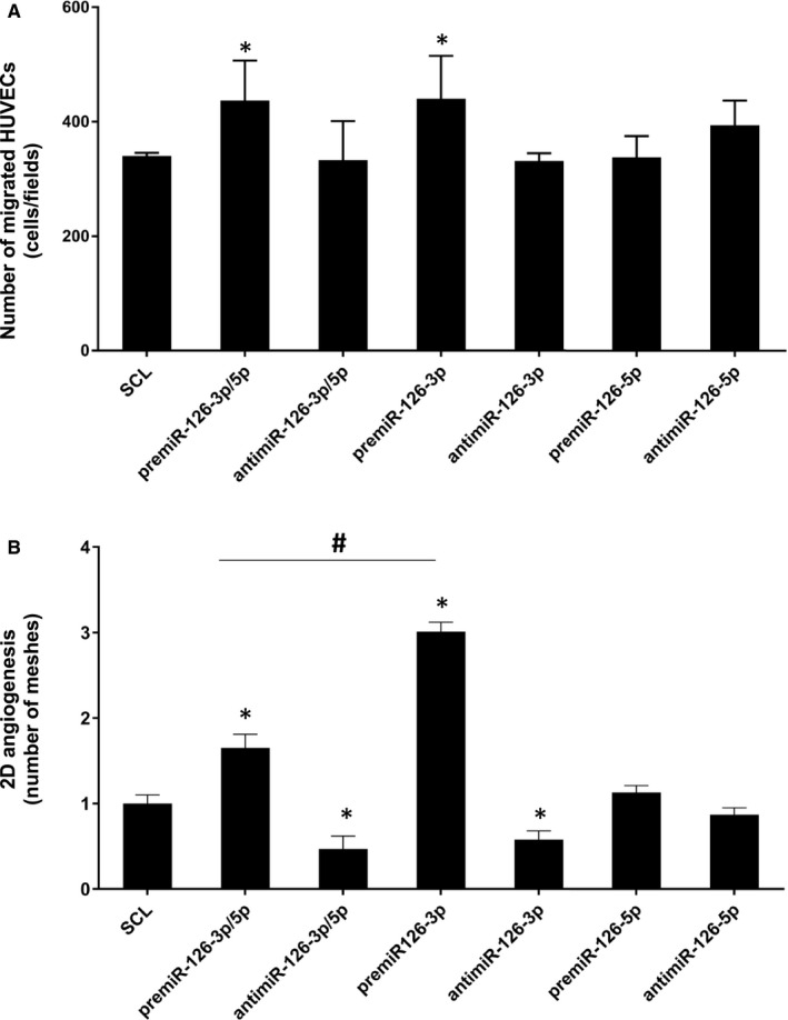
FIGURE 2.

Effect of miR‐126‐3p and miR‐126‐5p on HUVEC migration and vascular tubes formation. The miR‐126 was up‐ or down‐regulated by transfecting HUVEC with 20 nmol.L‐1 of premiR‐126 or inhibitors for 24 h. (A) Migration assay was performed using Boyden chamber. 5.104 transfected cells were seeded on the upper compartment during 24 h; the number of migrated cells was determined using phase contrast microscope. (B) Vascular tubes formation in 2D on Matrigel. 7500 transfected cells were deposited on the top of Matrigel and the tubular formation was studied after 6 h of incubation. The quantity of meshes was determined using phase contrast microscope and Archimed(TM) and Histolab(TM) software. For each assay, three independent experiments were performed. *p <.05 vs SCL; # p <.01 premiR‐126‐3p/5p vs premiR‐126‐3p
