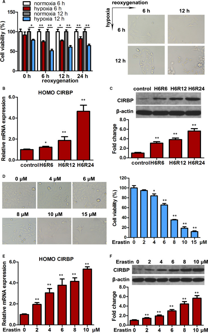
FIGURE 1.

CIRBP expression is increased under hypoxia/reoxygenation (HR) and during ferroptosis in HK2 cells. A, HK2 cells subjected to normoxia and hypoxia for 6/12 h were then cultured for 24 h, and cell viability was measured. Representative images are shown (right panel). B, Relative mRNA expression of CIRBP detected by qRT‐PCR in HK2 cells cultured under HR. C, CIRBP protein levels in the indicated cells were assessed by western blotting. Representative blot (upper panel) and quantification (lower panel) are shown. β‐actin was used as loading control. D, Cell viability of HK2 cells treated with erastin (0‐15 μmol/L) for 24 h. Representative images are shown (left panel). E, Relative mRNA expression of CIRBP detected by qRT‐PCR in HK2 cells treated with erastin (0‐10 μmol/L) for 24 h. F, CIRBP protein levels in the indicated cells were assessed by western blotting, and representative blots (upper panel) and quantification (lower panel) are shown. Values are expressed as the mean ± SD. n = 3, * P < .05, ** P < .01
