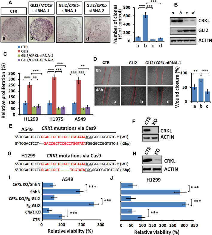
FIGURE 3.

CRKL is critical for GLI2‐driven cell proliferation and migration. A, Clone numbers of A549 cells after indicated treatments. Quantification analyses were shown on right. B, Western blot analysis of A549 cells treated with indicated siRNAs. ACTIN acts as a loading control. C, MTT analyses of indicated LUAD cells under indicated transfections for 48 hours. Notably, GLI2 promoted NSCLC cells proliferation, which was counteracted by CRKL knockdown. D, Wound healing assays of A549 cells under indicated treatments. Quantification of wound closure at indicated time points was shown on right. E, Alignment of Sanger sequencing results of PCR amplicons from A549 cells. SgRNA targets were highlighted in red and the PAM sequence was underlined. F, Western blot analysis of CRKL expression of wild‐type (WT) and knockout (KO) A549 cells. G, Alignment of Sanger sequencing results of PCR amplicons from H1299 cells. H, Western blot analysis of CRKL expression of wild‐type (WT) and knockout (KO) H1299 cells. I, J, MTT results of WT or CRKL KO A549 and H1299 cells under indicated treatments. Notably, knockout of CRKL decreased cell proliferation and blunted GLI2‐driven cell proliferation. All values are mean ± SD (n = 3, **P < .01 and ***P < .001)
