Fig. 3.
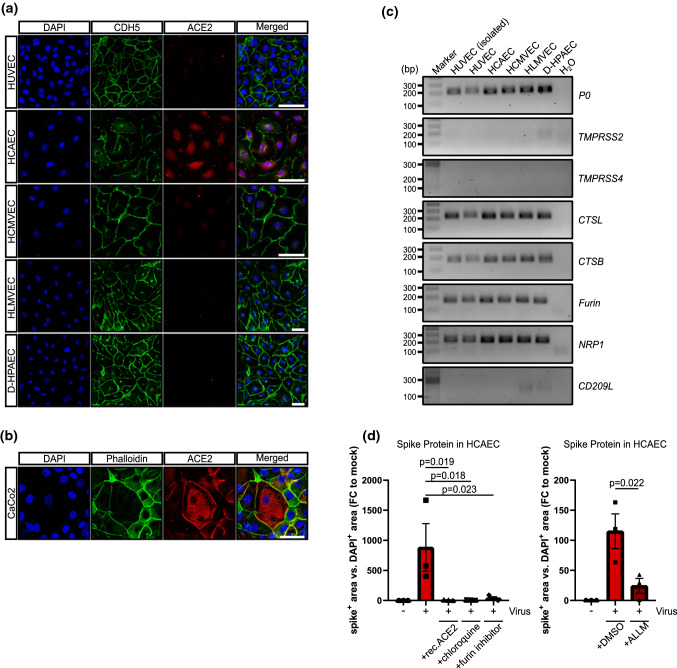
SARS-CoV-2 receptor expression in different endothelial cell cultures. a, b HUVEC, HCMVEC, HCAEC, HLMVEC, D-HPAEC and CaCo2 cells were seeded at 80% confluence and were stained 1 day after against ACE2 (red). DAPI (blue) and CDH5/phalloidin (green) served as counter staining. Scale bars = 50 µm. c RT-PCR products of TMPRSS2, TMPRSS4, CTSL, CTSB, FURIN, NRP1 and CD209L in cultured endothelial cells and freshly isolated HUVEC. PCR products were run on a 1.5% agarose gel in 1 × TAE buffer. d Spike protein quantification in HCAEC, 3 days after infection. Cells were incubated with 10 µM chloroquine, 1 µM (in DMSO) cathepsin inhibitor N-Acetyl-l-leucyl-l-leucyl-l-methional (ALLM) and 20 µM FURIN-inhibitor I. 5 µg/ml of human recombinant ACE2 was mixed with the virus and incubated for 30 min, prior to infection. Cells were fixed and stained against viral spike protein. Experiments were conducted in triplicate. Data are shown as mean and error bars indicate SEM. Data were statistically assessed using a one-way ANOVA test with a post hoc Dunnett’s test

