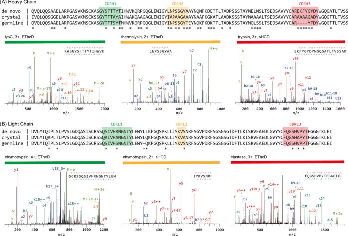
Figure 2.
Mass spectrometry-based de novo sequence of the mouse monoclonal anti-FLAG-M2 antibody. The variable regions of the heavy (A) and light chains (B) are shown. The MS-based sequence is shown alongside the previously published sequence in the crystal structure of the Fab (PDB ID: 2G60) and germline sequence (IMGT-DomainGapAlign; IGHV1-04/IGHJ2; IGKV1-117/IGKJ1). Differential residues are highlighted by asterisks (*). Exemplary MS/MS spectra in support of the assigned sequences are shown below the alignments with protease, precursor charge state, and fragmentation type indicated. Peptide sequence and fragment coverage are indicated on top of the spectra, with b/c ions indicated in blue/teal and y/z ions in red/orange. The same coloring is used to annotate peaks in the spectra, with additional peaks such as intact/charge reduced precursors, neutral losses, and immonium ions indicated in green. Note that to prevent overlapping peak labels, only a subset of successfully matched peaks is annotated.

