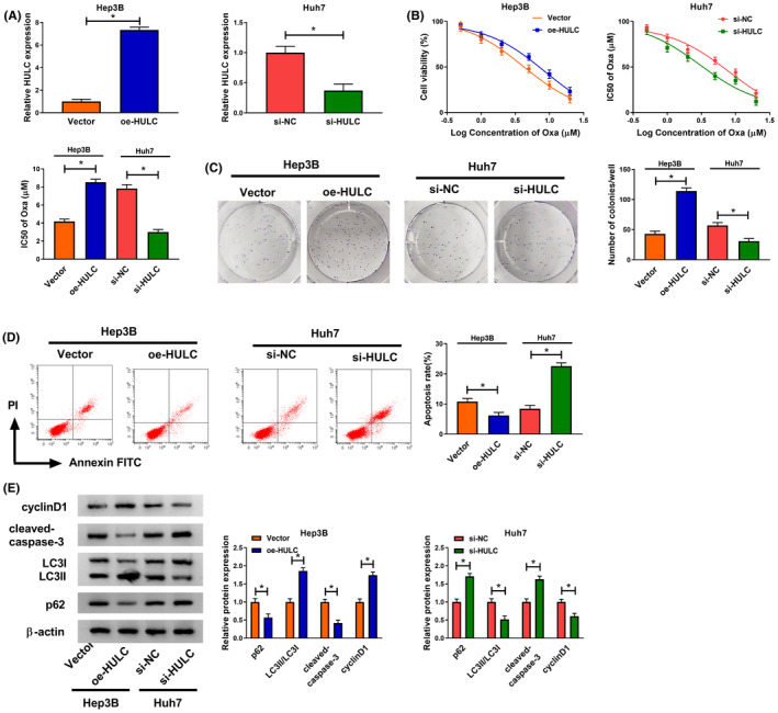
FIGURE 2.

HULC promoted the progression of HCC cells and chemosensitivity of Oxa. (A) Transfection efficiency of oe‐HULC in Hep3B cells and transfection efficiency of si‐HULC in Huh7 cells were detected by qRT‐PCR (t‐test). (B) Cell viability and IC50 of Oxa value were measured by CCK‐8 assay in Hep3B cells transfected with Vector or oe‐HULC or Huh7 cells transfected with si‐NC or si‐HULC before treatment with different concentrations of Oxa (0, 0.5, 1, 2, 5, 10, and 20 μM) (t‐test). (C–E) Hep3B cells were transfected with Vector or oe‐HULC and Huh7 cells were transfected with si‐NC or si‐HULC and then these cells were treated with Oxa (6 μM). (C) Colony formation assay was utilized to determine the number of colonies (t‐test). (D) Cell apoptosis was determined using flow cytometry analysis (t‐test). (E) Western blot assay was applied to determine the protein levels of cyclinD1 cleaved‐caspase‐3, LC3I/II, and p62 (t‐test). *p < .05. CCK‐8, Cell Counting Kit‐8; HCC, hepatocellular carcinoma; HULC, highly upregulated in liver cancer; LC3, light Chain 3; NC, negative control; qRT‐PCR, quantitative real‐time polymerase chain reaction
