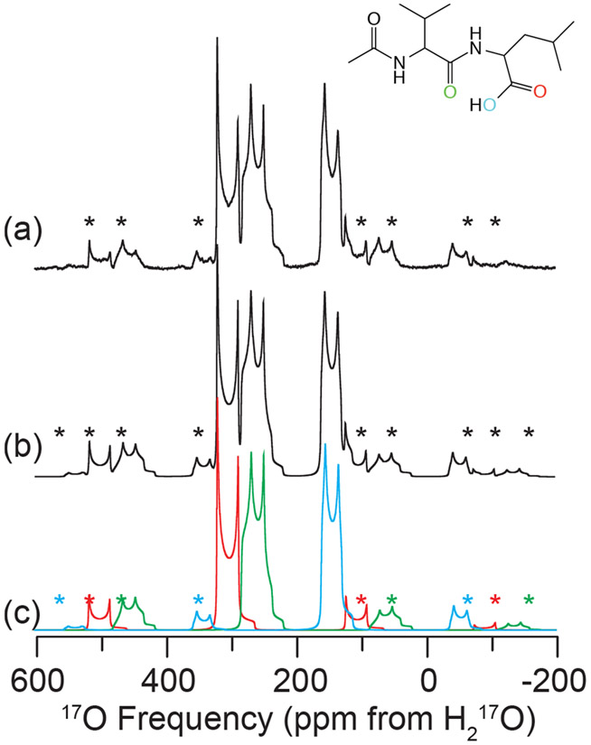
Figure 2.
Experimental 17O MAS NMR of N-Ac-VL (a), full simulation of MAS NMR spectrum (b) and simulation of each individual oxygen environment (c) at 21.1 T (ω0H/2π = 900 MHz). Line structure is shown in the inset indicating the 17O enriched sites: CO (red), NCO (green) and COH (blue). Spectra were acquired with ωR/2π = 24 kHz, spinning sidebands are noted by asterisks (*). NMR parameters used in spectral simulations are given in Table 4.

